Updated 26th of February 2021
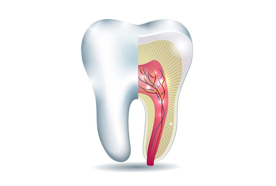
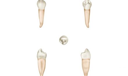
Cementum is a very thin and calcified layer of specialized tissue that covers the surface of the root of a tooth, firmly interlocked with the dentin. It ensures the attachment of the teeth to the alveolar bone by anchoring to the periodontal ligament.
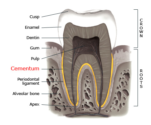
Cementum is part of the periodontium tissue complex which surrounds and supports teeth.
Structure
Cementum is a mineralized connective tissue similar to bone except that it does not contain blood vessels. It has a mineral part of which the major component is hydroxyapatite (calcium phosphate mineral) and an organic matrix which consists mainly of collagen.
The cells that form cementum are called cementoblasts. The produced cementum is slightly softer than dentin, which itself is softer than enamel.
Cementoenamel junction
CEJ, or cementoenamel junction, is a tooth area often talked about in dentistry. It refers to the cervical line of a tooth, where the cementum joins the enamel. This anatomically visible structure is sometimes called the neck of a tooth.
The CEJ is usually the place where healthy gums attaches to a tooth. If gingival recession occurs, the cementoenamel junction can be visually revealed, and is a sign of unhealthy teeth and gums.
There are three possible ways of how the cementum meets the enamel at the CEJ, and all three can be present in the same set of teeth, and even on the same tooth:
- The cementum overlaps the enamel (60–65% of teeth).
- The cementum and enamel border each other (30% of teeth).
- There is a space left between the enamel and cementum, where the underlying dentin is exposed (5-10% of teeth). This condition can make teeth sensitive to liquids and to temperature changes in the mouth.
Dentinocemental junction
The DCJ is not a clearly defined junction, as the tissues it separates, the cementum and the dentin, both have common histological and embryological backgrounds. It does not have a clinical significance, but is referred to the place on the dentin layer where cementum is located and starts its connection to the dentin.
Functions of the cementum
The cementum has three major functions that provide health and integrity to teeth.
Firstly, cementum gives connection to the periodontal ligament through components called Sharpey’s fibres. This enables teeth to be attached to the alveolar bone.
Secondly, cementum participates in maintaining the periodontal ligament space, which therefore ensures integrity of the periodontium (surrounding gum tissues) of a tooth.
Thirdly, cementum is a medium that repairs the root surface of a tooth if damage occurs.
Diseases of the cementum
Cementum is a less mineralized tissue than dentin and enamel, and may be subject to some diseases.
Hypercementosis is a situation where excessive thick cementum is formed around a root. This condition may affect a single tooth or multiple teeth. It can be associated with excessive forces from teeth grinding and clenching; in these cases we would observe wear on the enamel of those teeth. Hypercementosis can also be associated with Paget’s disease, a hereditary condition where, among other observations, there is generalized hypercementosis.
Cementicles are small globular masses of calcified cementum. Their size is usually less than 0.5 mm in diameter. They are either found within the cementum or in the periodontal ligament. Cementicles are thought to be the result of calcification due to cellular degeneration, but the reason why this takes place is unclear. Cementicles have no clinical significance, unless they are exposed to the oral environment, where they can cause accumulation of dental plaque, which may lead to tooth cavities or periodontal disease.
If cementum is exposed in a gingival recession condition, it may wear out more rapidly because of its low mineral content. This will expose the underlying dentin which may also wear out (abrasion), and can lead to tooth sensitivity. This condition makes a tooth more susceptible to dental caries at its exposed root surfaces.
References
- Yamamoto T, Hasegawa T, Yamamoto T, Hongo H, Amizuka N. (Histology of human cementum: Its structure, function, and development). Japanese Dental Science Review. 2016 Aug; 52(3): 63–74.
- Daly C G, J.B.Kieser JB, Corbet E F, Seymour J. (Cementum involved in periodontal disease: a review of its features and clinical management). Journal of Dentistry. Volume 7, Issue 3, September 1979, Pages 185-193.
- Pinheiro BC, Pinheiro TN, Capelozza AL, Consolaro A. (A scanning electron microscopic study of hypercementosis). Journal of Applied Oral Science Nov-Dec 2008;16(6):380-4.
- Periobasics.com, (Cementum).
- ScienceDirect, (Cementum).
- Wikipedia, (Cementum).
The information above should be used as a reference only. Any medical decision should not be taken before consulting a health care professional.